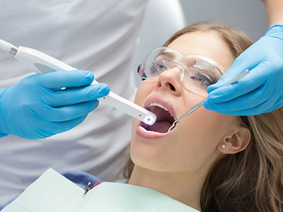
An intraoral camera is a compact, pen-sized imaging tool that captures high-resolution, full-color photographs of the inside of the mouth. Lightweight and maneuverable, these cameras provide a detailed, magnified view of teeth, gums, and other soft tissues, then display those images on a nearby monitor in real time. For patients, seeing a crisp, close-up image of a problem area makes oral conditions easier to understand and prepares them to take an active role in care decisions.
Intraoral cameras use tiny lenses and integrated lighting to illuminate and photograph areas that are difficult to see with the naked eye. The device’s tip is designed to reach around corners and into tight spaces, allowing clinicians to photograph individual tooth surfaces, restorations, and soft tissue with clarity. Modern systems produce images that are sharp enough for diagnostic evaluation, yet simple enough to be reviewed comfortably on a chairside display.
These cameras typically connect directly to a computer or imaging system, where images can be enhanced, annotated, and stored. Many practices integrate intraoral images with their electronic health records, which helps maintain a comprehensive visual history of a patient’s oral health. Because the images are digital, they can be quickly retrieved during follow-up visits or used as a visual reference while explaining treatment options.
Importantly, capturing an image with an intraoral camera is non-invasive and painless. Unlike radiographs, which show internal structures, intraoral photography records surface-level detail in full color, making it especially useful for assessing enamel wear, fractures, early-stage decay, soft-tissue abnormalities, and the condition of existing restorations.
Seeing is understanding. For many patients, an image of their own teeth bridges the gap between clinical terminology and real-world concerns. An intraoral camera equips patients with a clear visual reference, enabling more informed conversations about oral health. When clinicians can point to a photograph on the screen, patients often grasp the nature and urgency of an issue much faster than when relying on verbal descriptions alone.
Chairside images also support shared decision-making. When patients can compare current photographs with previous visits, they gain perspective on small changes that might otherwise go unnoticed. This transparency builds trust and encourages proactive care—patients are more likely to follow preventive recommendations or accept necessary treatments when they understand the “why” behind them.
For those who experience dental anxiety, intraoral imaging can actually reduce uncertainty. Visual feedback demystifies what’s happening inside the mouth and clarifies the steps clinicians propose to address any problems. The result is a calmer, more collaborative patient experience rooted in clear information rather than conjecture.
Clinicians use intraoral images as a complementary diagnostic tool alongside clinical exam findings and radiographs. A high-quality photograph can reveal subtle enamel defects, hairline fractures, margin discrepancies around crowns or fillings, and areas of early decay. These surface-level cues often guide the clinician toward more focused evaluations with other diagnostic modalities when necessary.
When planning restorative or cosmetic procedures, intraoral photography helps document the starting point and guide precise treatment choices. Detailed images enable better shade matching, margin assessment, and communication with laboratory technicians who fabricate crowns, veneers, or prosthetics. Intraoral images act as an objective visual record that supports predictable clinical outcomes.
In addition, images aid in monitoring periodontal and soft-tissue health. Photographs taken over time can show progression or improvement of inflammation, recession, or tissue healing after treatment. This visual timeline helps clinicians tailor follow-up care and reinforces the clinical rationale for maintenance or intervention strategies.
Digital intraoral images are easily archived as part of a patient’s permanent record. Keeping a well-organized library of photographs enhances continuity of care; clinicians can review visual history at any appointment to evaluate how an issue has evolved. These images streamline charting and reduce ambiguity during case reviews or long-term treatment planning.
Sharing images with specialists or a dental laboratory becomes efficient and precise with digital files. When collaborating on complex restorative work, an annotated photograph communicates details that are difficult to convey in words alone, such as exact margin locations, surface texture, and shade considerations. This visual collaboration improves coordination and reduces the likelihood of misinterpretation.
Many modern offices incorporate intraoral imaging into broader digital workflows, pairing photographs with intraoral scans, radiographs, and three-dimensional imaging. This combined dataset supports comprehensive treatment planning, whether the objective is conservative restoration, periodontal therapy, or full-mouth rehabilitation. The interoperability of these technologies enhances accuracy and speeds clinical decision-making.
If an intraoral camera is used during an appointment, the process is straightforward and comfortable. After explaining the purpose, your clinician will gently guide the camera along the surface of the teeth and tissues while you remain seated in the dental chair. The device’s small size makes it easy to maneuver; most patients feel nothing more than a light touch as images are captured.
Images appear instantly on a monitor so both patient and clinician can review them together. Your care team may highlight areas of concern, point out changes since your last visit, or demonstrate how recommended treatments will address specific problems. These conversations are framed around the images, which help turn abstract concepts into tangible visuals that are easy to follow.
Privacy and records management are part of the process: captured images are stored securely within the practice’s patient record system and accessed only by authorized personnel involved in your care. If an image needs to be shared with another clinician for consultation, your treatment team will manage that exchange as part of coordinated care planning.
Because intraoral photography is an everyday diagnostic tool, there’s no extra preparation required on your part. Whether you’re visiting for a routine checkup or discussing a proposed procedure, the chairside images simply make communication clearer and care more precise.
Intraoral cameras are an example of how digital tools can improve both the technical quality of care and the patient experience. By making oral conditions visible and understandable, they support better decisions, stronger communication, and more consistent long-term outcomes. If you’d like to learn more about how our team uses intraoral imaging to support diagnosis and treatment, please contact us for more information.


An intraoral camera is a compact, pen-sized imaging device that captures high-resolution, full-color photographs of the inside of the mouth. It uses a small lens and integrated lighting to illuminate tooth surfaces, restorations, and soft tissues while producing a magnified image in real time. The images appear on a chairside monitor so patients and clinicians can review findings together.
The camera’s slender tip is designed to reach around corners and into tight spaces that are difficult to see with the naked eye. Modern systems connect directly to a computer or imaging platform, allowing clinicians to enhance, annotate, and store images for clinical use. Because the images are digital, they can be retrieved quickly during follow-up visits to compare changes over time.
Chairside intraoral imaging bridges the gap between clinical terminology and what patients actually see in their mouths, making conditions easier to understand. When a clinician can point to a photograph on the screen, patients often grasp the nature and urgency of an issue more quickly than with verbal descriptions alone. This visual context supports informed, shared decision-making about care options.
Seeing clear images of their own teeth also helps patients recognize subtle changes that might otherwise go unnoticed, which encourages proactive preventive measures. Visual feedback can reduce anxiety by demystifying dental findings and clarifying recommended steps. Overall, chairside imaging fosters transparency, trust, and better adherence to treatment and maintenance plans.
Clinicians use intraoral photographs as a complementary diagnostic tool alongside clinical examinations and radiographs. High-quality images can reveal surface-level issues such as enamel defects, hairline fractures, margin discrepancies around restorations, and early-stage decay that guide further evaluation. Photographs often direct clinicians to focus imaging or testing on specific areas of concern.
In restorative and cosmetic cases, intraoral images document the starting condition and assist with shade matching, margin assessment, and communication with laboratory technicians. Annotated images provide objective visual records that support predictable outcomes and reduce ambiguity during case planning. The result is more precise treatment selection and clearer expectations for the patient.
Yes, intraoral images are typically stored as part of a patient’s digital record and organized for easy retrieval during future visits. These images become part of the permanent chart and are treated like other clinical documentation, helping clinicians track progression, treatment results, and healing. Access to the images is restricted to authorized members of the dental team who are involved in your care.
When images need to be shared with a specialist or a dental laboratory for consultation or fabrication, the exchange is managed securely and only with appropriate consent. Secure storage and controlled access help protect patient privacy while enabling efficient collaboration. Patients can request copies of their images or have them transferred as needed for continuity of care.
Intraoral photography is non-invasive, painless, and involves no ionizing radiation, making it a safe complement to standard dental imaging. Most patients feel only a light touch as the small camera is guided gently around the teeth and soft tissues while seated in the dental chair. The procedure is quick and typically causes no discomfort or recovery time.
Offices follow infection-control protocols such as barrier sleeves or disposable covers for the camera tip and routine device disinfection between patients. These measures ensure patient safety and maintain clinical hygiene standards. If you have concerns about sensitivity or gag reflex, discuss them with your clinician so they can adapt the technique for your comfort.
Intraoral cameras capture full-color surface images that show enamel texture, fractures, restoration margins, and soft-tissue appearance, while radiographs reveal internal structures such as tooth roots, bone levels, and hidden decay. Each modality provides distinct and complementary information important to a comprehensive evaluation. Clinicians often use both tools in combination to form a complete diagnostic picture.
Photographs are especially useful for documenting visible conditions and communicating findings to patients or laboratories, whereas radiographs are essential for assessing hidden pathology and bone health. Using both images together improves diagnostic accuracy and supports more informed treatment planning. Your clinician will recommend the appropriate imaging based on the clinical question at hand.
Yes, intraoral photography is an effective way to document and monitor periodontal and soft-tissue conditions over time. Sequential photographs can show changes in inflammation, gingival recession, tissue healing after treatment, or the progression of lesions that warrant further evaluation. These visual records help clinicians tailor maintenance intervals and verify the effectiveness of therapies.
When compared side by side, images make small but clinically significant changes easier to detect than memory or written notes alone. Photographs also facilitate communication about homecare needs and motivate patients to follow recommended hygiene protocols. Regular imaging as part of periodontal care enhances long-term tracking and clinical decision-making.
Annotated intraoral photographs convey details that are difficult to describe in words alone, such as exact margin locations, surface texture, and shade nuances. Sharing high-quality images with a dental laboratory or a specialist reduces misunderstandings and helps achieve restorations and treatment outcomes that match clinical intent. Visual information improves coordination for complex restorative or cosmetic cases.
Digital image files can be transmitted quickly and integrated into collaborative workflows, shortening turnaround times and reducing the potential for errors. Clear photographs supplement written prescriptions and measurements, giving technicians a precise visual reference. The result is better alignment between the clinician’s objectives and the final prosthetic or restorative work.
No special preparation is required for intraoral photography; the process is designed to be simple and unobtrusive. Most patients sit comfortably in the dental chair while the clinician briefly guides the camera around areas of interest to capture the necessary images. Because the procedure is quick and noninvasive, it integrates seamlessly into routine exams and treatment visits.
If you prefer, brushing your teeth before an appointment can reduce surface debris and improve image clarity, but it is not mandatory. If you have strong gag sensitivity or other concerns, tell your dental team so they can adjust technique or timing for greater comfort. Otherwise, simply arriving for your visit is all that’s needed.
At Whitesburg Dental Design, intraoral camera images are combined with other digital records—such as intraoral scans and radiographs—to create a comprehensive visual dataset for diagnosis and treatment planning. Integrating these images into the patient record supports precise restorative work, improves laboratory communication, and streamlines case coordination. The digital workflow helps the care team make faster, more informed decisions while keeping patients involved in the process.
Captured images are annotated and archived as part of each patient’s chart so clinicians can compare visits and document treatment outcomes over time. This interoperability enhances continuity of care for routine maintenance, surgical planning, or full-mouth rehabilitation. Patients benefit from clearer explanations and a transparent treatment process supported by objective visual evidence.
Ready to schedule your next dental appointment or have questions about our services?
Contacting Whitesburg Dental Design is easy! Our friendly staff is available to assist you with scheduling appointments, answering inquiries about treatment options, and addressing any concerns you may have. Whether you prefer to give us a call, send us an email, or fill out our convenient online contact form, we're here to help. Don't wait to take the first step towards achieving the smile of your dreams – reach out to us today and discover the difference personalized dental care can make.