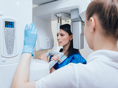
Digital radiography replaces traditional x-ray film with electronic sensors and computer imaging to capture detailed pictures of your teeth, jaws, and surrounding structures. When a sensor is placed in the mouth and exposed to a controlled x-ray beam, it converts the incoming radiation into a digital image that appears almost instantly on a monitor. This immediate feedback allows the clinician to confirm image quality before the patient leaves the chair, reducing the need for repeat exposures.
There are a few common types of digital sensors—direct sensors that transmit images immediately to the computer and plate systems that are scanned afterward—but they all serve the same purpose: to produce clear diagnostic images quickly and consistently. The software used with these systems can adjust contrast, zoom in on areas of interest, and measure structures, giving clinicians flexible tools to evaluate oral health with greater precision.
For patients, the most noticeable difference is speed. What once required film development and waiting now happens in seconds, enabling faster diagnosis and clearer communication between the dentist and the patient. That speed also supports more efficient appointments and helps clinicians make timely decisions about treatment plans.
One of the primary benefits of modern digital radiography is its ability to produce diagnostic images using significantly less radiation than traditional film techniques. Advances in sensor sensitivity and image-processing algorithms mean clinicians can obtain high-quality images at lower dose levels, adhering to accepted safety standards and principles such as ALARA (As Low As Reasonably Achievable).
Lower exposure is especially important for patients who require periodic monitoring or children, who are more sensitive to ionizing radiation. Because digital systems capture usable images more consistently, there is less chance of needing repeat shots due to under- or overexposure. When images can be reviewed immediately, the clinician can retake a single, properly exposed image right away if needed.
Beyond dose reduction, many modern practices use shielding and up-to-date x-ray units that further minimize risk. Digital radiography is one component of a comprehensive safety protocol designed to protect patients while delivering the diagnostic detail clinicians need to provide informed care.
Digital images often have higher contrast and resolution than conventional film, and digital tools enhance diagnostic capabilities. Software can highlight subtle differences in density, enlarge suspicious areas without losing clarity, and perform measurements that support accurate assessments of tooth structure, root health, and bone levels. These enhancements help clinicians detect problems earlier—such as small cavities between teeth or the early stages of bone loss—when treatment is typically simpler and less invasive.
Another advantage is the ability to compare current and past images side-by-side on screen. This comparative view helps track changes over time, monitor healing after procedures, and evaluate the effectiveness of ongoing treatments. Digital radiography integrates easily with other imaging technologies—such as intraoral cameras and cone-beam CT scans—helping create a complete diagnostic picture when complex issues arise.
Because image quality directly impacts clinical confidence, many dentists rely on digital radiography as a cornerstone of modern diagnostic care. It supports better treatment planning, clearer documentation, and more predictable outcomes for restorative, endodontic, and periodontal therapies.
Digital radiography improves efficiency across the entire patient experience. Images transfer instantly into the patient’s electronic record, eliminating the delays and storage demands associated with film. This seamless workflow shortens appointment times and allows treatment discussions to happen in real time, with images available for immediate reference.
Instant digital files are also simple to share securely with specialists, labs, or referring providers when collaborative care is necessary. That ease of sharing accelerates consultations and supports coordinated treatment plans, which is particularly helpful for cases that require orthodontic, surgical, or prosthetic input. Sending a high-quality digital image takes moments and preserves the full diagnostic value of the file.
From an operational perspective, going digital reduces physical storage needs and removes chemical waste from the office environment. Many practices find that modern digital systems not only speed up clinical processes but also support better recordkeeping and long-term case management.
For many patients, the experience of digital radiography is noticeably more comfortable than older film methods. Sensors are slim and come in sizes appropriate for different mouths; capturing an image is fast, and the team can confirm that the picture is usable before the appointment progresses. Because images appear immediately on the screen, dentists can walk patients through what they see, explaining findings with visual context rather than abstract descriptions.
Switching to digital often shortens appointment length since there is no development time and fewer repeat exposures. That convenience is especially valuable during restorative visits, where a quick image can confirm the fit of a restoration, the success of root canal therapy, or the condition of surrounding bone without scheduling a separate visit.
Patients should also expect that their digital images will be managed securely within the practice’s electronic record system. Care teams follow privacy protocols to protect health information, and images are available for future reference, making longitudinal care and follow-up more straightforward for both clinician and patient.
When a higher level of imaging is needed—such as three-dimensional views for implant planning—the dental team may recommend supplemental scans that complement two-dimensional digital radiographs. Together, these tools provide a comprehensive approach to diagnosis and treatment tailored to each patient’s needs.
High-quality imaging is central to accurate diagnosis, effective treatment, and clear communication. Digital radiography brings technical benefits that translate into tangible improvements in patient care: lower radiation exposure, faster and more comfortable appointments, and images that support earlier detection of issues. These advantages help clinicians deliver conservative, evidence-based treatments when they will be most effective.
At the office of Whitesburg Dental Design, adopting advanced imaging technologies reflects a broader commitment to efficient, patient-centered care. Technology does not replace clinical judgment; instead, it amplifies it—helping clinicians see more, explain more clearly, and plan with greater confidence.
Whether you are coming in for a routine exam or a more complex procedure, understanding how digital radiography fits into your care can help you feel more informed and involved. If you have questions about imaging protocols, safety measures, or what to expect during an x-ray appointment, please contact us for more information.
To learn more about how digital radiography supports timely, accurate dental care, or to discuss imaging as part of your treatment plan, contact us for more information.


Digital radiography uses electronic sensors and computer imaging to capture dental images instead of film, producing pictures almost instantly for immediate review. Sensors placed in the mouth convert x-ray energy into a digital file that appears on a monitor, allowing clinicians to confirm image quality before the patient leaves the chair. This instant feedback reduces the need for repeat exposures and eliminates film development time.
Unlike traditional film, digital systems include software tools that adjust contrast, zoom in on areas of interest, and take measurements that support diagnosis. The result is a faster workflow and clearer visual communication between clinician and patient, which helps guide treatment decisions during the same appointment.
Modern digital sensors are more sensitive to x-rays than film, so clinicians can obtain diagnostic images at lower dose levels while still maintaining image clarity. This adherence to the ALARA principle (As Low As Reasonably Achievable) means fewer repeat images and reduced cumulative exposure, which is especially important for children and patients who need frequent monitoring. Using up-to-date x-ray units and shielding further minimizes any unnecessary dose.
Because images appear immediately on screen, the operator can confirm whether an image is diagnostically acceptable and retake it once if necessary rather than exposing the patient to multiple repeated attempts. Overall, the combination of sensor sensitivity, software optimization, and modern equipment contributes to safer imaging without sacrificing diagnostic detail.
Dental practices commonly use two main intraoral sensor types: direct digital sensors that transmit images to a computer in real time, and phosphor plate systems that are scanned after exposure to produce a digital image. Direct sensors are known for immediate display and consistent image quality, while plate systems offer a thinner, flexible option that some patients find more comfortable. Extraoral digital detectors are used for panoramic and cephalometric images when a broader view of the jaws is needed.
These sensors vary by size and design to accommodate different mouths and clinical needs, and many integrate seamlessly with imaging software to allow contrast adjustments and measurements. For three-dimensional planning or complex anatomy, clinicians may recommend cone-beam CT scans as a complementary modality rather than a replacement for routine two-dimensional digital radiographs.
High-contrast, high-resolution digital images make it easier to spot subtle changes in tooth structure, small interproximal cavities, and early signs of bone loss than many traditional films. Software tools let clinicians enlarge areas of concern and adjust image parameters to highlight differences in density, supporting more accurate and earlier diagnosis. Early detection often leads to simpler, less invasive treatments and better long-term outcomes.
Digital records also enable side-by-side comparisons of current and prior images, which helps clinicians monitor progression or healing over time. This longitudinal view is particularly valuable for periodontal monitoring, evaluating endodontic success, and tracking the stability of restorations or implants.
Digital sensors are generally thinner and more flexible than traditional film holders, which many patients find more comfortable during image capture. Because images display instantly, clinicians can complete diagnostic checks and discuss findings during the same visit, reducing the need for follow-up appointments solely for imaging. The faster workflow also shortens overall chair time and helps keep visits on schedule.
Immediate visualization on-screen allows dentists to explain conditions visually, improving patient understanding and involvement in care decisions. Eliminating chemical processing from the office reduces unpleasant odors and environmental waste, contributing to a more pleasant experience for patients and staff alike.
Yes, digital radiographs are straightforward to share securely with specialists, referring providers, or dental laboratories, preserving full diagnostic quality during transfer. Files can be exported in standard formats and sent through encrypted communication channels or uploaded to secure portals approved by the practice. This rapid exchange accelerates consultations and supports coordinated care when multidisciplinary input is needed.
Because the images remain digital, clinicians can include annotation or measurement data that helps receiving providers assess the case more efficiently. Secure sharing reduces delays associated with physical film and ensures that collaborating clinicians have access to the same high-resolution images for treatment planning.
Digital images are typically stored in the practice's electronic record system and remain available for future comparisons, treatment planning, and follow-up care. Secure storage supports long-term case management by enabling clinicians to track healing, evaluate treatment response, and document changes over time without repeated baseline exposures. Access to prior images also streamlines coordination with specialists and supports continuity of care.
The practice follows privacy protocols to protect health information and manages image access according to professional and legal standards. If you have questions about how your images are stored or who may access them, the care team can explain the office's procedures for safeguarding records.
Three-dimensional imaging, such as cone-beam computed tomography (CBCT), is recommended when volumetric detail is required for complex cases—examples include implant planning, evaluation of impacted teeth, assessment of jaw pathology, or advanced endodontic diagnostics. CBCT provides depth and spatial relationships that two-dimensional radiographs cannot, improving surgical planning and risk assessment in anatomically challenging situations. Clinicians weigh the diagnostic benefits against radiation dose and use 3D imaging only when it will materially influence treatment decisions.
For most routine exams and restorative checks, two-dimensional digital radiographs provide adequate diagnostic information with lower exposure. When a clinician recommends supplemental 3D imaging, they will explain the reason, what information the scan will provide, and how it will inform the proposed treatment plan.
Preparation for dental x-rays is minimal: patients should inform the dental team if they are pregnant or have any health concerns that affect imaging, and remove jewelry or removable dental appliances if requested. During the procedure, a lead apron or thyroid collar may be used for added protection, and the clinician will position a sensor or plate in the mouth for a few seconds while the exposure is made. The process is quick and generally well tolerated by both adults and children.
After the exposure, images appear on the monitor almost immediately so the dentist can review them with you and answer questions while you are still in the chair. If additional views are needed, the team can take them right away, which helps avoid scheduling separate visits and keeps care efficient.
High-quality imaging supports accurate diagnosis, precise treatment planning, and clearer communication between clinicians and patients, all of which contribute to better outcomes and more predictable care. Digital radiography enables dentists to detect issues earlier, monitor changes over time, and tailor treatments based on detailed visual information rather than assumption. These capabilities reduce uncertainty and help clinicians choose conservative, evidence-based approaches when appropriate.
At Whitesburg Dental Design, incorporating advanced imaging into practice workflows reflects a commitment to efficient, patient-centered dentistry and informed decision-making. Technology enhances clinical judgment by providing clearer data, and when combined with professional expertise it helps deliver safer, more effective care.

Ready to schedule your next dental appointment or have questions about our services?
Contacting Whitesburg Dental Design is easy! Our friendly staff is available to assist you with scheduling appointments, answering inquiries about treatment options, and addressing any concerns you may have. Whether you prefer to give us a call, send us an email, or fill out our convenient online contact form, we're here to help. Don't wait to take the first step towards achieving the smile of your dreams – reach out to us today and discover the difference personalized dental care can make.