Modern dental care relies on clear information. Cone-beam computed tomography (CBCT) gives dentists a three-dimensional view of the teeth, jaws, and surrounding structures that traditional X-rays can’t match. When used thoughtfully, CBCT helps clinicians diagnose complex problems, plan procedures with confidence, and reduce guesswork—improving outcomes and patient comfort.
At the office of Whitesburg Dental Design, we incorporate CBCT into cases where added detail matters most. Our goal is to use this technology selectively and responsibly to support diagnoses and treatment plans that are safer, more predictable, and better tailored to each patient’s needs.
CBCT produces volumetric images that show anatomy from multiple angles rather than a single flat view. That three-dimensional perspective helps clinicians inspect tooth roots, bone height and density, the shape of the jaw, nerve pathways, and sinus anatomy in a single dataset. This breadth of information can expose issues that would otherwise be missed on two‑dimensional films.
Beyond teeth and bone, CBCT can clarify conditions affecting adjacent structures—temporomandibular joints, impacted teeth, suspected root fractures, and non-odontogenic sources of pain. Because the scan captures fine spatial relationships, it supports more accurate diagnoses when anatomy is complex or symptoms are ambiguous.
Interpreting CBCT volumes requires trained eyes. Our team reviews the full dataset to identify clinically relevant findings and to distinguish routine variations from signs that require intervention or referral. That careful interpretation is what turns detailed images into practical treatment decisions.
One of CBCT’s most valuable roles is in pre-procedural planning. For dental implants, the scan reveals bone quantity and quality and maps vital structures so implant position can be planned precisely. This reduces intraoperative uncertainty and helps ensure implants are placed in the optimal prosthetic position for function and aesthetics.
CBCT also enhances planning for extractions of impacted teeth, complex endodontic cases, and reconstructive procedures. By simulating the surgical view ahead of time, clinicians can anticipate potential challenges and select the most conservative, efficient approach.
In many cases, the CBCT dataset is used to design surgical guides, mill custom components, or collaborate with dental laboratories. Those digital workflows convert diagnostic detail into practical tools that streamline treatment and improve predictability from initial consult to final restoration.
Patient comfort and safety are central when using any radiographic technology. Modern CBCT units capture the needed volume quickly, often in a single rotation, which shortens scan time and minimizes motion artifacts. The result is a more comfortable experience compared with lengthy imaging sessions.
Exposure is closely managed through targeted fields of view and contemporary low-dose protocols. Rather than scanning more anatomy than necessary, clinicians select the smallest volume that answers the clinical question—an approach that balances diagnostic benefit with responsible radiation stewardship.
Before any scan, we discuss the purpose of the CBCT, what the patient can expect during the brief appointment, and how the images will be used in their care. That transparency helps patients feel informed and reassured about the role of imaging in their treatment.
CBCT is most powerful when it’s part of an integrated digital workflow. When combined with intraoral scans and digital design tools, the three-dimensional data guides restorative planning, implant placement, and fabrication of precise surgical guides or restorations. This integration reduces manual steps and improves alignment between surgical and prosthetic goals.
For example, merging a CBCT scan with a digital impression allows the team to position an implant to meet both anatomical constraints and the desired final appearance of the tooth. That forward-thinking coordination minimizes compromises down the line and helps deliver restorations that function well and look natural.
The ability to share digital files with specialists and dental labs also enhances collaboration. Detailed CBCT datasets provide partners with the same clear reference points, which speeds decision-making and reduces the likelihood of miscommunication during multi-disciplinary care.
CBCT is a targeted diagnostic tool used when two-dimensional imaging is insufficient or when three-dimensional detail directly affects a clinical decision. Typical indications include complex implant planning, assessment of impacted or unusually oriented teeth, evaluation of suspected root fractures, and investigation of unexplained facial pain or pathology.
Not every dental visit requires a CBCT scan. Routine hygiene checks and simple restorative needs are typically managed with standard radiographs and clinical examination. We recommend CBCT only when the additional anatomic information will alter the course of care.
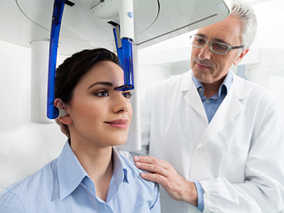
If a CBCT is recommended, the appointment is straightforward: the patient is seated or standing, positioned for the selected field of view, and the scan is completed in moments. Afterward, the clinician reviews the images and discusses findings and treatment options with the patient, ensuring a clear plan is in place.
At Whitesburg Dental Design, we use CBCT thoughtfully as part of an evidence-based approach: selecting scans when they add diagnostic value, interpreting results with clinical context, and integrating findings into treatment plans designed around each patient’s goals.
In summary, CBCT is a powerful diagnostic resource when applied selectively and interpreted by experienced clinicians. It provides a clearer view of dental and facial anatomy, supports more precise treatment planning, and integrates seamlessly with modern digital workflows to improve predictability and patient care.
For more information about how CBCT might be used in your care, please contact us to discuss your needs and learn whether 3D imaging is appropriate for your situation.


Cone-beam computed tomography (CBCT) is a three-dimensional imaging technique that captures a volumetric dataset of the teeth, jaws and surrounding structures in a single rotation. The unit projects a cone-shaped X-ray beam and a specialized detector to record multiple images, which are reconstructed into a 3D model clinicians can view in axial, coronal and sagittal planes. This volumetric view reveals spatial relationships and fine anatomic detail that cannot be appreciated on conventional two-dimensional radiographs.
At the office of Whitesburg Dental Design we use CBCT selectively to answer specific diagnostic questions and to support precise treatment planning. Because the dataset contains multiplanar views and cross-sectional slices, it becomes a practical diagnostic resource for complex cases where depth and spatial orientation matter.
Traditional intraoral and panoramic X-rays provide two-dimensional representations of dental anatomy, which can compress overlapping structures and hide critical details. CBCT produces true three-dimensional images that preserve depth and spatial relationships, allowing clinicians to view anatomy from multiple angles and to isolate structures without superimposition. This difference is especially important when evaluating root anatomy, bone volume, nerve position and the relationship of teeth to adjacent sinuses or anatomic landmarks.
While CBCT offers richer diagnostic information, it is not a replacement for routine 2D radiographs in every situation; instead, it complements conventional imaging when additional anatomic detail will affect clinical decisions. Clinicians weigh the expected diagnostic benefit against radiation exposure and select the modality that best answers the clinical question.
CBCT is recommended when three-dimensional detail will change diagnosis or treatment—for example, complex implant planning, assessment of impacted or aberrantly positioned teeth, investigation of suspected root fractures, and evaluation of unexplained facial pain or pathology. It is also valuable for pre-surgical planning in reconstructive cases, endodontic assessment of complex canal anatomy, and when pathology is suspected that extends beyond the teeth. These indications reflect situations where 2D imaging may be insufficient or misleading.
Not every dental visit requires CBCT; routine exams and many restorative procedures are adequately managed with standard radiographs and clinical evaluation. The decision to scan is individualized and based on clinical signs, symptoms and whether the 3D information will materially influence the proposed care plan.
For implant planning, CBCT provides accurate information about bone height, width and density as well as the precise location of vital structures such as the inferior alveolar nerve and the maxillary sinuses. This allows the clinician to select appropriate implant length and diameter, avoid critical anatomy, and identify the need for grafting or other preparatory procedures. The 3D dataset reduces intraoperative uncertainty and helps establish an optimal prosthetic-driven position for the planned restoration.
CBCT data can also be used to design fabrication of surgical guides, which transfer the virtual plan to the clinical setting and improve placement accuracy. Integrating CBCT with digital impressions and CAD/CAM workflows helps align surgical and prosthetic goals, enhancing functional and aesthetic outcomes while reducing the likelihood of unplanned adjustments during surgery.
CBCT uses ionizing radiation, so safety and justification are essential. Modern CBCT systems and contemporary protocols emphasize dose reduction through limited fields of view, optimized exposure settings and faster acquisitions; clinicians select the smallest volume that answers the clinical question to minimize exposure. When used appropriately, the diagnostic benefits often outweigh the risks, particularly in complex cases where 3D information alters treatment decisions.
Patient-specific factors are considered before scanning, and alternative imaging options are evaluated when appropriate. Pregnant patients and those with special considerations are managed according to professional guidelines, and clinicians discuss the rationale and safety measures with patients before proceeding.
A CBCT appointment is typically brief and noninvasive: the patient is seated or standing and positioned so the area of interest is centered in the unit, and the scan is completed in a single rotation that lasts only a few seconds to a minute. Because the capture is fast, motion artifacts are reduced and overall patient comfort is generally high compared with longer imaging procedures. No contrast agents or injections are required for standard dental CBCT scans.
After the scan, the clinician reviews the images and discusses the findings and recommended next steps with the patient, explaining how the 3D data informs diagnosis and treatment planning. The practice will also handle any necessary referrals or collaboration with specialists based on the imaging results.
CBCT images should be interpreted by clinicians trained in evaluating three-dimensional dental and maxillofacial anatomy; this often includes general dentists with CBCT training, oral and maxillofacial surgeons, endodontists or oral radiologists depending on the clinical context. Skilled interpretation involves reviewing multiplanar reconstructions, volumetric renderings and cross-sectional slices to identify clinically relevant findings and to differentiate normal anatomic variants from pathology. When findings are complex or outside the clinician’s scope, referral to or consultation with a specialist or oral and maxillofacial radiologist is appropriate.
Interpretation guides treatment decisions such as implant placement, surgical approach, endodontic intervention or the need for further diagnostic testing. At Whitesburg Dental Design our clinicians integrate CBCT findings with clinical examination and other records to create clear, evidence-based treatment plans tailored to each patient’s needs.
Yes. CBCT captures the surrounding facial structures and is useful for assessing the maxillary sinuses, temporomandibular joints (TMJ), airway anatomy and bony pathology of the jaws. It can identify sinus disease that relates to dental infections, condylar bone changes in TMJ disorders and osseous lesions that might require further evaluation. The ability to visualize adjacent anatomy is one reason CBCT is valuable when symptoms are atypical or when dental sources of pain are uncertain.
However, CBCT primarily shows bony and hard-tissue detail; soft-tissue conditions may require complementary imaging modalities or specialist evaluation. Clinicians interpret CBCT findings in the broader clinical context and recommend additional tests or referrals when soft-tissue assessment or medical imaging is indicated.
CBCT is central to modern digital workflows because the volumetric dataset can be merged with intraoral scans and prosthetic designs to create a coordinated, prosthetically driven plan. This integration enables virtual implant positioning that accounts for both anatomic constraints and the desired final restoration, improving alignment between surgery and prosthetics. Digital planning files can be exported to design and mill patient-specific surgical guides, which help transfer the virtual plan to the clinical setting with greater precision.
Sharing CBCT-derived files with dental laboratories and specialists streamlines collaboration and reduces the risk of miscommunication during multi-disciplinary care. The digital workflow often shortens treatment timelines and increases predictability by minimizing manual steps and ensuring all team members work from the same detailed reference data.
Preparation for a CBCT scan is minimal. Patients should remove jewelry, eyeglasses, removable dental appliances and any metal objects in the head and neck area that could cause artifacts in the images, and they should bring prior radiographs or relevant records if requested by the clinician. Patients should also inform the team if they are pregnant or suspect they might be, as additional precautions or alternatives may be considered.
The appointment itself is short and requires no special dietary or activity restrictions; after the scan the clinician will review the images and explain the next steps. If further consultation or specialist involvement is needed based on the findings, the practice will coordinate appropriate follow-up to ensure a clear plan of care.
Ready to schedule your next dental appointment or have questions about our services?
Contacting Whitesburg Dental Design is easy! Our friendly staff is available to assist you with scheduling appointments, answering inquiries about treatment options, and addressing any concerns you may have. Whether you prefer to give us a call, send us an email, or fill out our convenient online contact form, we're here to help. Don't wait to take the first step towards achieving the smile of your dreams – reach out to us today and discover the difference personalized dental care can make.